Postoperative Urinary Retention Management: How Bladder Scanners Are Changing Surgical Recovery
Postoperative urinary retention management is one of the most overlooked aspects of surgical recovery, yet it directly impacts patient outcomes, hospital discharge timelines, and infection rates. When a patient cannot void after surgery, clinical teams face a critical decision. Do they catheterize and accept the associated risks, or do they monitor and wait? Without objective bladder volume data, that decision is often based on time elapsed and clinical intuition rather than evidence.
Portable bladder scanners have fundamentally changed this equation. A bladder scan after surgery gives clinicians a real time, non-invasive measurement of how much urine is in the bladder, transforming postoperative urinary retention from a clinical guessing game into a data driven management pathway. The result is fewer unnecessary catheterizations, earlier detection of true retention, faster discharge, and fewer hospital acquired infections.
This guide covers everything surgical teams, PACU nurses, and clinical administrators need to know about managing postoperative urinary retention with bladder scanning technology, including evidence-based protocols, catheterization thresholds, and practical implementation strategies. 🩺
Understanding Postoperative Urinary Retention
Postoperative urinary retention, commonly abbreviated as POUR, is the inability to urinate after a surgical procedure despite having a full bladder, in a patient without a prior history of urinary disorders. It is one of the most common postoperative complications and occurs across virtually all surgical specialties, though its incidence varies significantly depending on the type of surgery, the anesthetic technique used, and patient specific risk factors.
Reported incidence rates range from as low as 5 percent to as high as 70 percent depending on the surgical population studied and the diagnostic criteria used. Joint replacement surgery, gynecological procedures, anorectal surgery, and hernia repair are among the procedures most associated with POUR. Spinal and epidural anesthesia carry higher retention risk than general anesthesia, and opioid pain medications further increase that risk by suppressing detrusor muscle contractility.
Why Early Detection Matters
Delayed recognition and treatment of postoperative urinary retention can lead to significant complications. Bladder overdistension stretches the detrusor muscle beyond its functional capacity, which can result in long term bladder dysfunction, overflow incontinence, and the need for prolonged catheterization. Acute retention with high bladder volumes can trigger autonomic responses including hypertension, hypotension, vomiting, and cardiac arrhythmias, all of which complicate postoperative recovery.
Equally important, undetected retention that leads to catheterization carries its own set of risks. Every catheter insertion introduces the possibility of catheter associated urinary tract infection, urethral trauma, and patient discomfort. On the other hand, catheterizing a patient who does not actually have clinically significant retention is an unnecessary invasive procedure that exposes the patient to harm without benefit.
This is precisely why a bladder scan after surgery is so valuable. It provides the objective data needed to distinguish between patients who genuinely need intervention and those who simply need more time to void naturally. 📊
How Bladder Scanning Improves Postoperative Urinary Retention Management
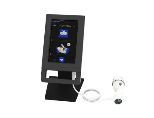
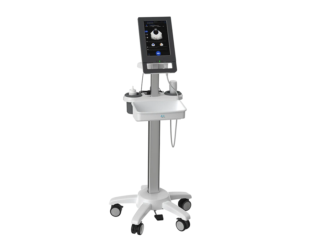
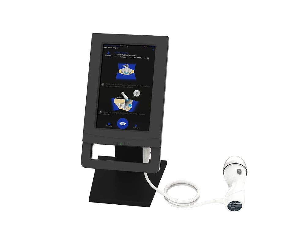
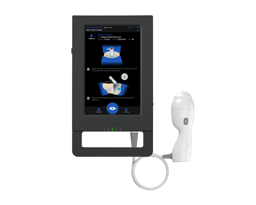
A portable 3D bladder scanner uses ultrasound technology to measure the volume of urine in the bladder non-invasively through the abdominal wall. The measurement takes less than three seconds, is painless, and can be performed by any trained member of the nursing staff without requiring a sonographer or physician.
In the context of postoperative urinary retention management, bladder scanning serves several critical functions. It provides an objective baseline for determining whether a patient’s bladder volume has reached a level that warrants intervention. It allows clinicians to distinguish between low volume states where the patient simply has not yet produced enough urine to void, and high-volume states where genuine retention is occurring. It supports the timing of intermittent catheterization by showing exactly when the bladder has reached a predefined threshold. And it provides post void residual data after a patient has attempted to urinate, confirming whether the bladder is emptying adequately.
Replacing Guesswork with Data
Before bladder scanning technology was widely available, clinical teams relied on time based triggers, abdominal palpation, and subjective patient assessment to manage postoperative voiding. A common practice was to catheterize any patient who had not voided within a set number of hours after surgery, regardless of their actual bladder volume. This approach led to significant over-catheterization because many of those patients simply did not yet have enough urine in their bladder to trigger the voiding reflex.
Research has demonstrated that time since last void is a poor predictor of bladder volume. Two patients may both be six hours post-surgery without voiding, but one may have only 150 mL in their bladder while the other has 600 mL. The appropriate clinical response for each patient is completely different, and only a bladder scan can reveal that distinction. Facilities that have transitioned from time-based protocols to volume-based protocols using bladder scanners consistently report reductions in catheterization rates without any increase in adverse events from missed retention.
Designing a Voiding Trial Protocol After Catheter Removal
For patients who had an indwelling catheter placed during surgery, the transition from catheterized to independent voiding is a critical moment. A well-designed voiding trial protocol after catheter removal ensures that patients are monitored appropriately during this transition and that retention is detected early without resorting to unnecessary re-catheterization.
Key Components of an Effective Voiding Trial Protocol
A voiding trial protocol after catheter removal should include several essential elements. The protocol should specify the timing of catheter removal, which in many evidence-based programs occur at a standardized interval after surgery, such as six hours postoperatively or on the morning of postoperative day one, depending on the surgical procedure and institutional practice.
After catheter removal, the protocol should define a monitoring window during which the patient is expected to void. A common timeframe is three to six hours. During this window, nursing staff should encourage the patient to attempt voiding, help with ambulation to the bathroom, and use non-pharmacological strategies such as running water and suprapubic warm compresses to stimulate the voiding reflex.
If the patient has not voided by the end of the monitoring window, a bladder scan should be performed to assess the actual bladder volume. This is the critical decision point. If the bladder volume is below a defined threshold, typically 300 to 400 mL depending on institutional protocol, the patient can continue to be monitored. If the volume exceeds the threshold, intermittent straight catheterization should be performed to relieve the retention, and the voiding trial should be restarted.
Post Void Residual Assessment
Once a patient does void after catheter removal, the job is not done. A post void residual measurement using the bladder scanner should be performed immediately after the patient urinates. This confirms that the bladder is emptying adequately. A post void residual volume of less than 200 mL is generally considered a successful voiding trial. Volumes between 200 and 400 mL may warrant continued monitoring, while volumes above 400 mL suggest incomplete emptying and may require further intervention.
The combination of timed monitoring, volume-based decision making, and post void residual assessment creates a comprehensive voiding trial protocol that balances patient safety with the goal of minimizing unnecessary invasive procedures. This approach has been shown to reduce re-catheterization rates, shorten hospital stays, and facilitate earlier discharge. 🏥
Bladder Scan Intervals and Catheterization Thresholds
One of the most common questions from clinical teams implementing bladder scanning protocols is how often to scan and at what volume to intervene. While institutional protocols vary, the evidence base provides clear guidance on effective scan intervals and catheterization thresholds for postoperative urinary retention management.
Recommended Scan Intervals
For patients in the post anesthesia care unit who have not voided, the first bladder scan should be performed when the patient reports symptoms of retention or when a defined time period has elapsed since surgery. Many protocols initiate scanning at three hours post-surgery or at the time of PACU discharge if the patient has not voided. Subsequent scans should be performed at regular intervals, typically every one to two hours, until the patient voids or until catheterization is indicated by the bladder volume.
For patients on the surgical ward who have had their catheter removed, scanning should begin if the patient has not voided within three to six hours of catheter removal. Repeat scanning every two hours after the initial scan provides ongoing monitoring without being excessively burdensome for nursing staff.
Evidence Based Catheterization Thresholds
The optimal catheterization threshold has been the subject of considerable clinical research. Traditional protocols often used thresholds of 300 to 400 mL, but more recent evidence suggests that higher thresholds may be safe and appropriate in many clinical scenarios. One large prospective study found that raising the catheterization threshold from 400 mL to 800 mL and implementing volume dependent scan intervals reduced the need for sterile intermittent catheterization by more than 65 percent without increasing catheterization rates on the ward or causing adverse events.
The current expert consensus, as reflected in guidelines published by SHEA and IDSA, recommends that clinical teams establish clear, evidence-based catheterization thresholds and communicate them consistently across all units. For most adult patients, a threshold of 400 to 500 mL with symptoms of retention, or 500 to 800 mL regardless of symptoms, represents a reasonable starting point. Institutions should adjust these thresholds based on their patient population, surgical mix, and quality improvement data.
Postoperative Urinary Retention Across Surgical Specialties
While the principles of postoperative urinary retention management are consistent across specialties, the specific risk profile and management considerations vary. Understanding these nuances helps clinical teams tailor their bladder scanning protocols for maximum effectiveness.
Orthopedic Surgery
Joint replacement surgery, particularly total hip and total knee arthroplasty, carries a significant risk of postoperative urinary retention. Contributing factors include spinal anesthesia, perioperative opioid use, older patient populations, and prolonged operative times. Many orthopedic centers have implemented routine bladder scanning protocols in the PACU and on the surgical floor, with good results. Selective scanning based on symptoms and inability to void within defined timeframes, rather than universal scanning of every patient, has been shown to reduce unnecessary interventions and improve discharge efficiency.
Gynecological and Obstetric Surgery
Urinary retention following gynecological procedures is common, particularly after pelvic surgery involving structures adjacent to the bladder and urethra. Procedures for uterine prolapse, ovarian cancer, and uterine cancer carry elevated retention risk due to surgical disruption of pelvic nerve pathways. Sling procedures for stress urinary incontinence, including TVT-O and TVT-S, also require careful postoperative bladder monitoring because over-correction can lead to voiding difficulty.
In obstetrics, postpartum urinary retention is an underrecognized complication that can occur after both vaginal delivery and cesarean section. Epidural anesthesia, prolonged labor, and instrumental delivery are risk factors. A bladder scan after delivery provides early detection and guides clinical decisions about whether to monitor or intervene. Routine postpartum bladder scanning has been adopted by many obstetric units as part of their standard recovery protocol.
Anorectal and Colorectal Surgery
Postoperative urinary retention rates after anorectal surgery are among the highest of any surgical specialty, with some studies reporting rates approaching 80 percent. The close anatomical relationship between the rectum and the lower urinary tract means that surgical site swelling, local anesthesia effects, and pain all contribute to voiding difficulty. Bladder scanning is essential in this population to grade the severity of retention and guide catheterization decisions that prevent secondary injury from bladder overdistension.
Hernia Repair and General Surgery
Inguinal hernia repair, particularly under spinal anesthesia, is associated with postoperative urinary retention in a meaningful percentage of patients. Laparoscopic abdominal surgery generally carries lower retention risk than open procedures, but patient age, opioid use, and intravenous fluid administration volumes all influence postoperative bladder function. A standardized approach to bladder scan after surgery in these populations ensures consistent detection and appropriate management regardless of the specific procedure performed.
Risk Factors for Postoperative Urinary Retention
Identifying patients at elevated risk for postoperative urinary retention before surgery allows clinical teams to plan proactive monitoring strategies. The major risk factors supported by the clinical evidence include the following. 📋
Patient age is a significant factor, with patients over 60 years old at substantially higher risk. Male sex is an independent risk factor, largely due to the contribution of prostatic hypertrophy to bladder outlet resistance. Pre-existing lower urinary tract symptoms, including urgency, frequency, hesitancy, and weak stream, indicate underlying bladder dysfunction that may be exacerbated by surgery and anesthesia.
The type of anesthesia plays a major role. Spinal and epidural anesthesia directly suppress the sacral nerve pathways that control bladder emptying, and the effects can persist for hours after the surgical procedure is complete. Opioid analgesics suppress detrusor contractility and reduce the urge to void. High intraoperative fluid volumes increase bladder filling rates in the immediate postoperative period, when the patient may not yet have recovered normal voiding sensation.
Surgical duration is also relevant, with procedures lasting longer than two hours associated with higher retention rates. And certain medications with anticholinergic properties, which are common in the perioperative setting, can further impair bladder function.
For patients with multiple risk factors, a proactive approach to postoperative urinary retention management that includes early and repeated bladder scanning is warranted to ensure timely detection and intervention.
Implementing Bladder Scanning in Your Surgical Recovery Program
Bringing bladder scanning technology into your surgical recovery workflow does not require a complicated implementation plan. The key steps are straightforward and can be accomplished in phases.
Step 1: Secure the Equipment
Purchase or lease portable bladder scanners and position them in the PACU, surgical floors, and any recovery areas where postoperative patients are managed. A 3D ultrasound bladder scanner with an accuracy of ±5 percent, a scan speed under three seconds, and an intuitive touchscreen interface ensures that frontline staff can use the device confidently with minimal training.
Step 2: Develop Your Protocol
Working with your surgical, nursing, anesthesia, and quality improvement teams, develop a written protocol for postoperative bladder management. The protocol should cover catheter removal timing, expected voiding timelines, bladder scan intervals, catheterization thresholds, post void residual assessment criteria, and documentation requirements. Adapt the protocol to the specific needs of your surgical population, but anchor it in the published evidence and national guidelines.
Step 3: Train Your Staff
Conduct hands on training sessions for all nursing staff who will be performing bladder scans. Training should cover device operation, proper probe placement, interpretation of results, and the decision-making framework outlined in your protocol. Most modern bladder scanners are designed for ease of use and can be learned quickly, but formal training ensures consistency and builds confidence.
Step 4: Launch, Monitor, and Refine
Begin using the protocol on a pilot unit, collect data on catheterization rates, voiding trial success rates, post void residual values, and CAUTI incidence, and use that data to refine your approach before scaling to additional units. Share your results with staff regularly to maintain engagement and demonstrate the impact of the program. Quality improvement is an iterative process, and even small refinements over time can yield significant gains in patient outcomes and operational efficiency. ✅
The Bottom Line for Surgical Teams
Postoperative urinary retention management is a clinical challenge that affects patients across every surgical specialty. When managed with time-based triggers and subjective assessment alone, it leads to over-catheterization, delayed discharge, and preventable healthcare associated infections. When managed with portable bladder scanning technology and evidence-based protocols, it becomes a controlled, data driven process that protects patients and improves outcomes.
A bladder scan after surgery takes less than three seconds, causes no discomfort, and gives the clinician the one piece of information they need most, how much urine is in the bladder. That single data point transforms the entire management pathway from reactive to proactive, from variable to standardized, and from invasive to non-invasive.
For surgical teams, PACU nurses, and clinical leaders who are committed to improving postoperative care, implementing a voiding trial protocol after catheter removal that includes routine bladder scanning is one of the highest impact, lowest barrier quality improvement initiatives available today.
To learn more about portable 3D bladder scanners that support postoperative urinary retention management and CAUTI prevention programs, contact our team for a clinical demonstration or volume pricing for your facility.