
High Frequency Ultrasound Skin Scanners – DUB Skin Scanner🔬🩺
High frequency ultrasound skin scanners provide advanced, non invasive imaging to support the detection and evaluation of skin cancer and a wide range of dermatologic conditions 🌿. Designed for clinical precision, these systems allow dermatologists and specialists to visualize skin structures in exceptional detail, supporting confident diagnosis, treatment planning, and ongoing patient monitoring.
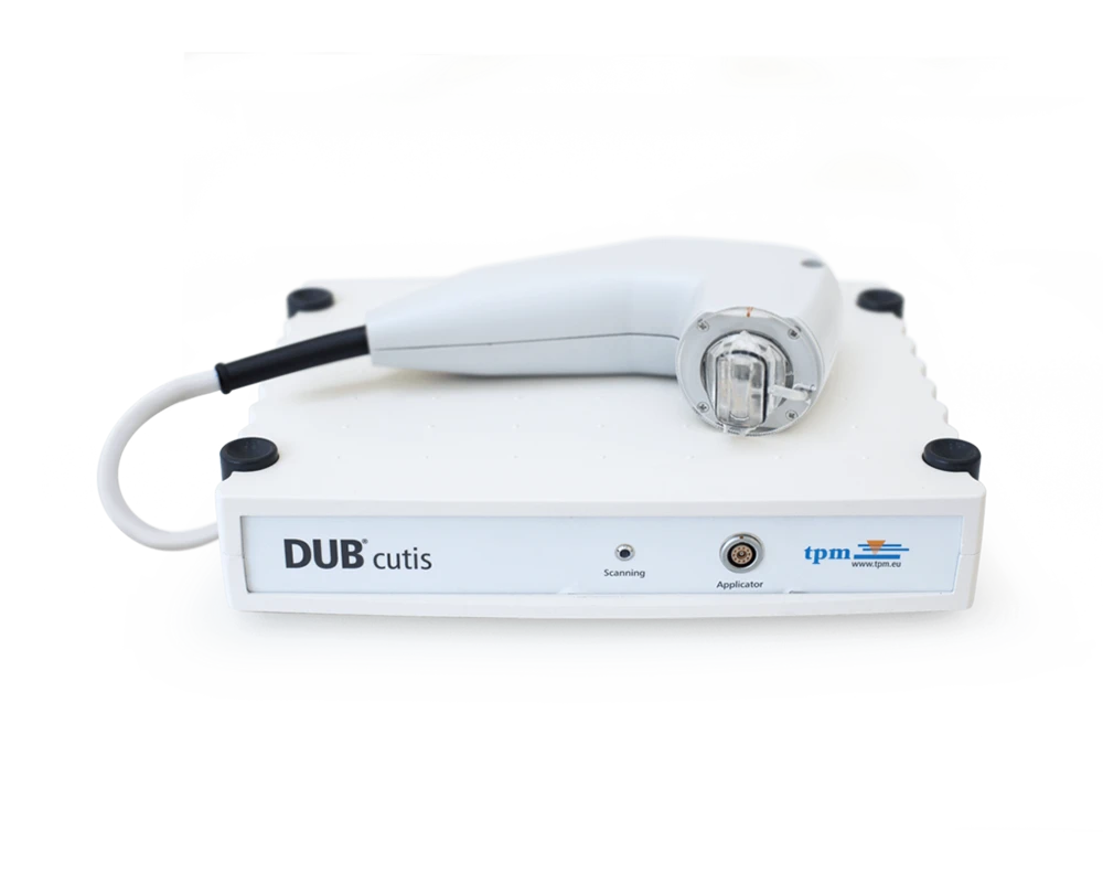
Our portfolio includes three distinct product types tailored to different clinical needs. The Dub Cutis offers reliable high frequency ultrasound imaging for routine dermatologic assessment, while the Dub Skin Scanner series expands capabilities across three models. The standard Skin Scanner delivers high quality imaging, the 75 MHz model provides enhanced resolution for superficial skin analysis, and the 100 MHz model offers the highest level of detail for advanced skin cancer detection and research focused applications 🧠📊.
As frequency increases, image clarity and structural detail improve, allowing clinicians to better assess lesion depth, margins, and tissue characteristics before intervention. Together, these high frequency ultrasound skin scanners support accurate, efficient, and patient focused dermatologic care in both clinical and research environments 🏥✨
High Frequency Ultrasound Skin Scanners for Advanced Dermatologic Imaging 🔬🩺
High frequency ultrasound skin scanners have become an essential component of modern dermatology, offering clinicians a powerful, non invasive way to visualize skin structures far beyond what is possible with the naked eye or dermoscopy alone 🌿. By using very high ultrasound frequencies, these systems generate detailed cross sectional images of the epidermis, dermis, and subcutaneous tissue, supporting accurate diagnosis, treatment planning, and follow up for a wide range of dermatologic conditions. In particular, high frequency ultrasound skin scanners play a growing role in the detection and evaluation of skin cancer, inflammatory skin disease, and cosmetic or procedural outcomes.
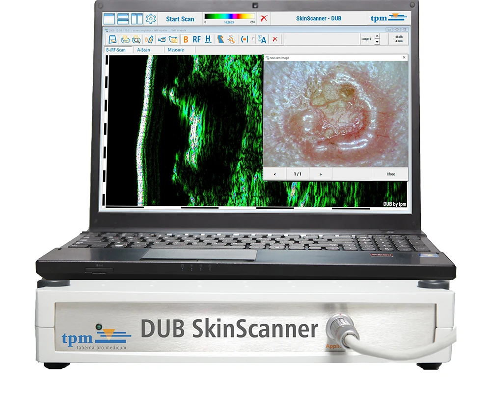
Within this category, the DUB product family includes three core solutions designed to meet different clinical needs and levels of diagnostic detail. These systems are the DUB Cutis, the DUB SkinScanner, DUB SkinScanner75, and the Dub SkinScanner100. Each platform is optimized for specific frequency ranges, allowing dermatologists to select the most appropriate high frequency ultrasound skin scanner based on the depth of tissue to be examined, the level of image resolution required, and the clinical question being addressed 🧠📊.
Together, these three systems form a comprehensive imaging ecosystem for dermatology, dermato oncology, aesthetic medicine, and clinical research. From routine skin assessment to ultra high resolution evaluation of suspicious lesions, high frequency ultrasound skin scanners enable clinicians to make better informed decisions while maintaining a patient friendly and non invasive diagnostic approach 🏥✨.
Dub SkinScanner Probes
DUB Cutis High Frequency Ultrasound Skin Scanners for Routine Dermatologic Assessment 🌱
The DUB Cutis is designed as an accessible yet highly capable high frequency ultrasound skin scanner for everyday dermatologic use. It is typically configured with probes operating in the lower high frequency range, which allows dermatologists to visualize superficial skin layers with clarity while maintaining sufficient penetration depth for routine assessment. This balance makes the DUB Cutis particularly suitable for general dermatology clinics, outpatient practices, and research settings where standardized skin measurements are required.
In clinical practice, dermatologists use the DUB Cutis high frequency ultrasound skin scanner to examine epidermal thickness, dermal structure, and the presence of focal lesions. This is especially valuable when assessing benign versus suspicious lesions, where changes in echogenicity and lesion boundaries can provide additional information beyond visual inspection 🔍. For patients with suspected basal cell carcinoma or squamous cell carcinoma, the DUB Cutis allows clinicians to estimate lesion depth and lateral extent, supporting early decision making regarding biopsy or excision.
Another important application of the DUB Cutis is the monitoring of chronic skin conditions such as psoriasis, eczema, or scleroderma. By using high frequency ultrasound skin scanning, clinicians can document changes in skin thickness and density over time, offering objective data to support treatment adjustments 📈. In cosmetic and aesthetic dermatology, the DUB Cutis is also used to evaluate skin hydration, dermal remodeling, and the effects of non invasive treatments, making it a versatile tool across multiple dermatologic disciplines.
DUB SkinScanner High Frequency Ultrasound Imaging for Enhanced Diagnostic Confidence 🔬
The DUB SkinScanner represents the next level of performance in high frequency ultrasound skin scanners, offering improved resolution and expanded clinical versatility. With support for higher frequency probes and advanced imaging modes, this system is well suited for dermatologists who require more detailed visualization of skin structures while still maintaining practical penetration depth for clinical use.
In the context of skin cancer diagnostics, the DUB SkinScanner high frequency ultrasound skin scanner enables more precise evaluation of lesion morphology. Dermatologists can assess lesion thickness, internal structure, and boundary definition, which is critical when planning surgical excision or monitoring lesion progression 🧠. This capability is particularly valuable for melanocytic lesions, where subtle differences in depth and echotexture can influence management decisions.
Beyond oncology, the DUB SkinScanner is widely used for lesion characterization and differential diagnosis. Conditions that may appear similar on the skin surface can present distinct ultrasound patterns when examined with high frequency ultrasound imaging. By adjusting probe frequency and imaging parameters, dermatologists can evaluate cystic versus solid lesions, inflammatory infiltrates, and vascular components with greater confidence.
The DUB SkinScanner also plays a significant role in treatment monitoring. Dermatologists frequently use high frequency ultrasound skin scanners to document therapeutic response in inflammatory skin diseases or to evaluate tissue changes following aesthetic procedures such as fillers, laser treatments, or skin tightening therapies 💉✨. The ability to capture repeatable, high quality images over time supports both clinical decision making and patient communication.
DUB SkinScanner75 Ultra High Frequency Ultrasound Skin Scanners for Maximum Detail 🧬
At the highest end of the imaging spectrum, the DUB SkinScanner75 is an ultra high frequency ultrasound skin scanner designed for clinicians who require the most detailed skin imaging available. Operating at frequencies up to 75 MHz, this system delivers exceptional resolution that allows visualization of fine micro structural details within the skin.
For dermatologists focused on skin cancer detection, the DUB SkinScanner75 provides unparalleled insight into lesion architecture. The ultra high frequency ultrasound images reveal subtle variations in tissue density and structure that can help distinguish malignant from benign lesions 🧠. This is particularly important in complex cases such as recurrent tumors, lentigo maligna, or lesions in cosmetically sensitive areas where precise margin assessment is essential.
The DUB SkinScanner75 is also widely used in academic and research environments, where ultra high resolution imaging is required to study skin physiology, aging processes, and treatment effects at a micro anatomical level. In aesthetic medicine, the system supports detailed evaluation of superficial skin layers, making it ideal for assessing fine lines, dermal thickness, and post treatment remodeling.
By allowing dermatologists to select different MHz ranges within a single platform, the DUB SkinScanner75 offers flexibility without compromising image quality. This makes it a powerful high frequency ultrasound skin scanner for advanced clinical practices seeking the highest diagnostic confidence 🔬📊.
DUB SkinScanner100 and Its Best Clinical Applications 🔬🩺
The DUB SkinScanner100 is designed for dermatologists who require the highest possible image resolution from a high frequency ultrasound skin scanner. Operating at 100 MHz, this system provides ultra high resolution imaging of superficial skin layers, allowing clinicians to visualize fine microstructural details with exceptional clarity. This makes the SkinScanner100 especially valuable in cases where precise assessment of lesion architecture and skin morphology is critical.
One of the primary clinical applications of the DUB SkinScanner100 is melanoma evaluation. The ultra high frequency imaging allows dermatologists to examine lesion thickness, internal structure, and margins with greater confidence, supporting earlier detection and more informed biopsy or excision planning 🧠. The detailed visualization helps differentiate subtle changes in pigmented lesions that may not be apparent through surface examination alone.
The SkinScanner100 is also well suited for assessing non melanoma skin cancers such as basal cell carcinoma and squamous cell carcinoma, particularly when lesions are superficial or located in cosmetically sensitive areas. By providing clear insight into tumor depth and boundary definition, the system supports precise surgical planning while helping preserve healthy tissue 🏥.
In addition, the DUB SkinScanner100 is widely used in inflammatory and cosmetic dermatology. Its high resolution imaging supports objective evaluation of conditions such as morphea, scleroderma, and chronic inflammatory dermatoses, as well as detailed assessment of skin remodeling following aesthetic treatments. This combination of precision and versatility makes the DUB SkinScanner100 a powerful tool for advanced dermatologic practice ✨
Using High Frequency Ultrasound Skin Scanners for Melanoma and Major Skin Conditions 🧠🩺
High frequency ultrasound skin scanners play an increasingly important role in the evaluation of melanoma by allowing dermatologists to visualize lesion structure beneath the skin surface in a non invasive way 🔬. When assessing suspected melanoma, ultrasound imaging helps determine lesion thickness, internal architecture, and boundary definition, which are critical factors in clinical decision making. By examining these characteristics before biopsy or surgical excision, clinicians can better plan treatment strategies and identify lesions that require urgent intervention.
Beyond melanoma, high frequency ultrasound skin scanners are widely used to evaluate other major skin cancers such as basal cell carcinoma and squamous cell carcinoma. These conditions often present with subtle surface features while extending deeper into the dermis. Ultrasound imaging allows dermatologists to assess tumor depth and lateral spread, supporting more accurate surgical planning and helping reduce the risk of incomplete excision 🏥. This capability is especially valuable in cosmetically sensitive areas where tissue preservation is essential.
High frequency ultrasound imaging is also highly effective in the assessment of inflammatory and structural skin conditions. Dermatologists use ultrasound scanners to examine diseases such as psoriasis, eczema, morphea, and hidradenitis suppurativa by visualizing changes in skin thickness, edema, and tissue density 📊. These insights support objective monitoring of disease progression and treatment response over time, offering measurable data beyond visual inspection alone.
In addition, these scanners are frequently applied to the evaluation of benign lesions, cysts, lipomas, and vascular abnormalities. By distinguishing between solid and fluid filled structures and analyzing echogenic patterns, dermatologists can refine differential diagnoses and reduce unnecessary invasive procedures 🌿. Across melanoma detection and a broad spectrum of dermatologic conditions, high frequency ultrasound skin scanners provide clinicians with deeper insight, improved diagnostic confidence, and enhanced patient care.
Clinical Use Cases and Procedure Based Applications for High Frequency Ultrasound Skin Scanners 🧠🏥
High frequency ultrasound skin scanners are increasingly integrated into dermatologic workflows because they provide actionable clinical information without exposing patients to radiation or invasive procedures. Across the DUB Cutis, DUB SkinScanner, and DUB SkinScanner75 platforms, three core use case categories consistently demonstrate the clinical value of this technology.
Skin cancer detection and tumor depth assessment represent one of the most important applications of high frequency ultrasound skin scanners. Dermatologists use ultrasound imaging to evaluate lesion thickness, internal structure, and margins before surgical intervention. This information supports better surgical planning, helping clinicians choose appropriate excision margins and reduce the likelihood of recurrence. High frequency ultrasound skin scanning also allows non invasive monitoring of suspicious lesions over time, supporting earlier intervention when changes are detected 🔍.
Lesion characterization and differential diagnosis form the second major use case. Many dermatologic conditions present with similar surface appearances but differ significantly in underlying structure. High frequency ultrasound skin scanners allow clinicians to differentiate cystic lesions from solid tumors, inflammatory conditions from fibrotic changes, and benign growths from malignant ones by analyzing echogenic patterns and tissue interfaces 🧬. This improves diagnostic accuracy and reduces unnecessary biopsies.
Monitoring therapeutic response and procedural outcomes is the third key application. Dermatologists increasingly rely on high frequency ultrasound skin scanners to track changes in skin structure following medical or aesthetic treatments. Whether assessing response to biologic therapy in inflammatory disease, evaluating scar remodeling, or documenting filler placement, ultrasound imaging provides objective, repeatable data that enhances clinical confidence and patient trust 📊✨.
Choosing the Right High Frequency Ultrasound Skin Scanners for Dermatologic Practice 🩺
Selecting the appropriate high frequency ultrasound skin scanners depend on the clinical focus, patient population, and diagnostic requirements of the practice. The DUB Cutis is well suited for routine dermatologic assessment, offering reliable imaging for general skin evaluation and follow up. The DUB SkinScanner provides enhanced resolution and flexibility, making it ideal for practices with a strong focus on lesion diagnostics and treatment monitoring. The DUB SkinScanner75 delivers the highest level of detail, supporting advanced skin cancer diagnostics, research, and specialized aesthetic applications.
By incorporating high frequency ultrasound skin scanners into daily practice, dermatologists gain access to a deeper understanding of skin pathology while maintaining a patient centered, non invasive diagnostic approach 🌿. These systems not only improve diagnostic accuracy but also enhance communication with patients by visualizing conditions in a clear and understandable way.
Advancing Dermatologic Care with High Frequency Ultrasound Skin Scanners 🚀
As dermatology continues to evolve toward precision medicine and minimally invasive diagnostics, high frequency ultrasound skin scanners play an increasingly central role. The DUB Cutis, DUB SkinScanner, and DUB SkinScanner75 together offer a scalable and future ready imaging solution that supports early detection, accurate diagnosis, and effective treatment planning across a wide spectrum of dermatologic conditions.
From everyday clinical assessment to ultra high resolution skin cancer evaluation, these high frequency ultrasound skin scanners empower clinicians with the imaging clarity and confidence needed to deliver high quality dermatologic care 🧠🏥.
Search Terms
dermatology ultrasound device, skin ultrasound imaging system, non invasive skin cancer imaging, dermatologic diagnostic ultrasound, cutaneous ultrasound imaging, ultrasound imaging for melanoma assessment, ultrasound for basal cell carcinoma evaluation, dermatology imaging equipment, skin lesion imaging technology, ultrasound for tumor margin assessment, dermatology ultrasound platform, clinical skin imaging system, ultrasound device for dermatologists, skin imaging ultrasound technology, high resolution dermatology ultrasound